<?xml version="1.0" encoding="UTF-8"?>
<?xml-stylesheet type="text/xsl" href="/stylesheets/feed.atom.xml" media="screen"?>
<feed xml:lang="en-US" xmlns="http://www.w3.org/2005/Atom">
  <id>tag:qbio.nd.edu,2005:/news</id>
  <link rel="alternate" type="text/html" href="https://qbio.nd.edu"/>
  <link rel="self" type="application/atom+xml" href="https://qbio.nd.edu/news.atom"/>
  <id>tag:qbio.nd.edu,2005:/latest</id>
  <title>Q-Bio@ND | News</title>
  <updated>2024-08-23T10:00:00-04:00</updated>
  <link rel="alternate" type="text/html" href="https://qbio.nd.edu/"/>
  <link rel="self" type="application/atom+xml" href="https://qbio.nd.edu//news.atom"/>
  <subtitle>QBio@ND, an interdisciplinary group at Notre Dame, brings together graduate students and faculty in Science and Engineering to advance quantitative biology.</subtitle>
  <entry>
    <id>tag:qbio.nd.edu,2005:News/165960</id>
    <published>2024-08-23T10:00:00-04:00</published>
    <updated>2024-08-30T14:54:57-04:00</updated>
    <link rel="alternate" type="text/html" href="https://qbio.nd.edu/news/piezo-proteins-sculptors-in-organ-growth/"/>
    <title>Piezo proteins, sculptors in organ growth</title>
    <summary type="text">
      <![CDATA[Butterfly wings, fish fins, and human limbs develop precisely and symmetrically. While genetics and chemical environment significantly influence their development, recent research has revealed that mechanical forces play a pivotal role as well. Piezo proteins have the unique ability to convert mechanical forces—such as the pressure and stretch of developing cells—into chemical signals. While these proteins have been previously shown to regulate blood pressure and sense pain, chemical and biomolecular engineers at the University of Notre Dame have demonstrated their crucial role in organ growth, regulating organ size, and the arrangement of cells in organ tissue. Their results were published in Cell Reports.]]>
    </summary>
    <content type="html">
      <![CDATA[<p>Butterfly wings, fish fins, and human limbs develop precisely and symmetrically. While genetics and chemical environment significantly influence their development, recent research has revealed that mechanical forces play a pivotal role as well.</p>
<p>Piezo proteins have the unique ability to convert mechanical forces—such as the pressure and stretch of developing cells—into chemical signals. While these proteins have been previously shown to regulate blood pressure and sense pain, chemical and biomolecular engineers at the University of Notre Dame have demonstrated their crucial role in organ growth, regulating organ size, and the arrangement of cells in organ tissue.</p>
<p>Their results were published in <a href="https://www.cell.com/cell-reports/pdf/S2211-1247(24)00726-5.pdf">Cell Reports</a>.</p>
<p>“Piezo acts like the thermostat in your house, it’s constantly measuring and adjusting the conditions of the cells,” said <strong><a href="https://engineering.nd.edu/faculty/jeremiah-zartman/">Jeremiah Zartman</a>,</strong> associate professor of chemical and biomolecular engineering at the University of Notre Dame.</p>
<section class="wp-block-uagb-columns uagb-columns__wrap uagb-columns__background-none uagb-columns__stack-tablet uagb-columns__valign-center uagb-columns__gap-10 align uagb-block-06412d20 uagb-columns__columns-2 uagb-columns__max_width-custom not-in-view in-view">
<div class="uagb-columns__inner-wrap uagb-columns__columns-2">
<div class="wp-block-uagb-column uagb-column__wrap uagb-column__background-undefined uagb-block-398051df">
<p>Fruit flies, with their fully sequenced genome, provided <a href="https://sites.nd.edu/zartmanlab/">Zartman’s lab</a> with a well-studied model for organ development that has many commonalities with organ growth in humans.</p>
<p>While it was known that Piezo proteins play a multifaceted role in cell growth and differentiation, the researchers were after a holistic picture of how this protein functioned on the larger scale of organ development.</p>
<p>On a cellular level, Piezo proteins join to create a gated channel into the cell membrane. Under mechanical tension, the channel’s gate opens, allowing calcium ions to flow in. The concentration of these ions cues the cell’s next step—proliferate, push other cells out, or undergo programmed cell death.</p>
<p>The team reverse engineered this signaling process, altering the levels of Piezo with drugs or genetic manipulations to better understand how these channels function. The resulting wing asymmetries, aberrant cell death, and changes in cellular proliferation highlighted the protein’s fundamental importance in organ development, even to non-adjacent tissues.</p>
<p>“It was a major surprise to us to find a protein, one that exists predominantly in the cell membrane, that could specifically control robustness, or precision,” said Zartman. “Piezo regulates how cells interact with each other, reach a certain size, stop growing, and it does this to ensure significant precision, there’s very little difference between one side of an organ and the other.”</p>
<p>Moving forward, Zartman said that his multi-institutional team will use mice and fish to explore how Piezo signals healthy cell development versus cancerous growth.</p>
<p>The team’s ongoing work is funded by the National Science Foundation’s (NSF) Emergent Mechanisms in Biology of Robustness, Integration &amp; Organization (EMBRIO) Institute and was supported by the NIH’s National Institute of General Medical Science (NIGMS) with early support from the NSF-Simons Center for Quantitative Biology Pilot program.</p>
</div>
</div>
</section>]]>
    </content>
    <link rel="enclosure" type="image/jpeg" href="https://qbio.nd.edu/assets/581522/zartman_tw.jpg" title="Fruit flies used in biomolecular research. Five vials of fruit flies inside with beige food on black lab benchtop"/>
    <author>
      <name>Karla Cruise</name>
    </author>
  </entry>
  <entry>
    <id>tag:qbio.nd.edu,2005:News/168301</id>
    <published>2024-06-04T11:00:00-04:00</published>
    <updated>2024-11-18T11:45:43-05:00</updated>
    <link rel="alternate" type="text/html" href="https://qbio.nd.edu/news/notre-dame-study-demonstrates-that-bacterial-biofilms-are-not-the-same-throughout-possibly-describing-one-reason-common-antibiotics-may-fail/"/>
    <title>Notre Dame study demonstrates that bacterial biofilms are not the same throughout, possibly describing one reason common antibiotics may fail</title>
    <summary type="text">
      <![CDATA[“We didn’t know how diverse the bacterial behavior was in such a small amount of space,” said Joshua Shrout, whose article was published in the Journal of Bacteriology.]]>
    </summary>
    <content type="html">
      <![CDATA[<p>The bacterium Pseudomonas aeruginosa is almost everywhere — in the soil, water, and vegetation. While it does not cause disease for most people, it can lead to serious disease or death for people with compromised immune systems.</p>
<p>The bacteria easily adhere to surfaces, creating structures called biofilms. <a href="https://engineering.nd.edu/faculty/joshua-shrout/">Joshua Shrout, professor in the Department of Civil and Environmental Engineering and Earth Sciences in the College of Engineering,</a> and collaborators have discovered that these biofilms are not the same throughout, and that substances called alkyl quinolones (AQs) are critical to this diversity.</p>
<p>“We didn’t know how diverse the bacterial behavior was in such a small amount of space,” said Shrout, <a href="https://journals.asm.org/doi/abs/10.1128/jb.00095-24?af=R">whose article</a> was published in the Journal of Bacteriology. “Understanding that if you treat it as homogenous, and it’s not, is one of the reasons classic antibiotics can fail: These bacteria are literally not behaving the same here as they are over there.”</p>
<figure class="image image-right"><img src="https://science.nd.edu/assets/570920/dcf.jpg" alt="Imaging of biofilm using several methods" width="600" height="648">
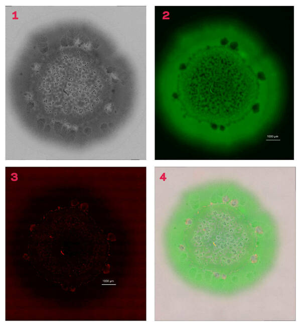
<figcaption>1. The image of a biofilm under light microscopy2. Live cells3. Dead cells 4. Merging of the entire series of images.</figcaption>
</figure>
<p>The research team began their study with what they thought was the simplest example of bacterial growth: a colony of cells on a petri plate. “It turns out this canonical example of microbiology isn’t basic or simple at all,” said Shrout, who is affiliated with the <a href="https://imaging.nd.edu/">Integrated Imaging Facility</a> and the <a href="https://precisionhealth.nd.edu/">Berthiaume Institute of Precision Health</a>.</p>
<p>Lead author Abigail Weaver, <a href="https://engineering.nd.edu/faculty/abigail-weaver/">assistant research professor in the Department of Civil and Environmental Engineering and Earth Sciences</a>, first noticed the presence or absence of large-scale structures within these bacterial colonies.</p>
<p>“The imaging that is used here is lightyears beyond what we could have done a decade ago, which is why no one has seen this diversity before,” Shrout said.</p>
<p>Weaver coordinated use of light microscopy and chemical imaging to collect a series of high-resolution pictures. When reassembled, the profiles showed unexpected variety within these colony biofilms — a variety that occurs behaviorally as well as spatially. This approach allows researchers to obtain different information about these biofilms with each layer they reassemble.</p>
<p>Pointing to an image of a biofilm, Shrout described areas of green, which are the live cells, and said that the image was overlaid with a standard light image. In addition to the cells, there are visible places, when viewed under high magnification, where there are gaps in the biofilm. To determine whether those blank areas have dead cells or simply did not have cells, researchers overlaid another layer that displayed the dead cells in red.</p>
<p>“In most of the cases where it looked like cells were missing, there were actually dead cells,” Shrout said. “It seems that they killed themselves off.”</p>
<p>Graduate students Jin Jia and Allison R. Cutri, in the laboratory of <a href="https://chemistry.nd.edu/people/paul-bohn/">Paul Bohn</a>, the Arthur J. Schmitt Professor in the Department of Chemistry and Biochemistry, used chemical imaging to confirm the presence of AQs in the same areas where dead cells were present.</p>
<p>To further understand the heterogeneous nature of the biofilm, the researchers combined high magnification light and chemical imaging to match up spatial features with chemical features. They determined four distinct regions in these colony biofilms that are differentiated by the presence or absences of large-scale structures, and studied the importance of how AQs are associated with aggregation formation and cell death.</p>
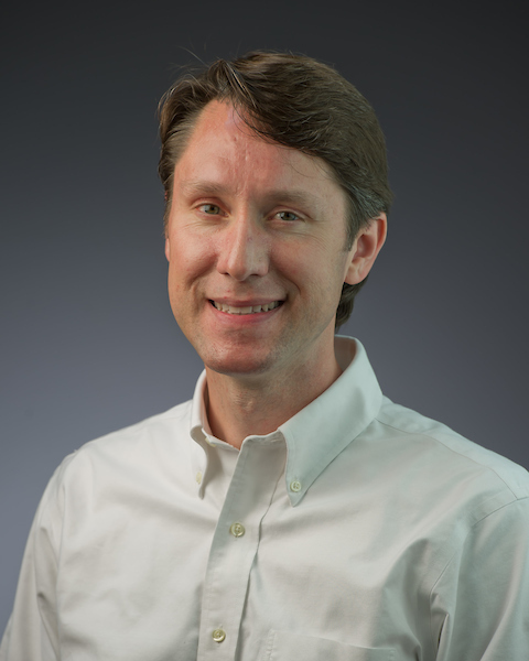
<figure class="image image-left"><img src="https://science.nd.edu/assets/570923/joshua_shrout.jpg" alt="Joshua Shrout" width="480" height="600">
<figcaption>Joshua Shrout</figcaption>
</figure>
<p>The AQs made by the bacteria seemed critical to making the diverse areas of the biofilms, so to test that hypothesis, researchers deleted one gene in the Pseudomonas aeruginosa that prevented that bacteria from making any AQs.</p>
<p>“We still see some differences in spatial heterogeneity with the AQ-deficient strain. But we don't see any of these pockets, where we see these aggregates or huge gaps of cells missing, right? It just looks pretty clean,” said Shrout, who is also a concurrent professor in the <a href="https://biology.nd.edu/">Department of Biological Sciences</a> in the College of Science, as he showed the images of the biofilm after gene deletion. “So that allows us to pretty definitively conclude these AQs are important for when we see all this crazy heterogeneity, including the aggregates and the dead cells.”</p>
<p>Though researchers knew AQs were important to the development of biofilms, the new knowledge about the extensive diversity within the biofilm, in such a small space, might aid in either the development or application of new drug classes to treat Pseudomonas aeruginosa.</p>
<p>“If you’re treating this infection as if it’s homogeneous, and it’s not, then you don’t get the result you expect. But you shouldn’t be surprised, right?” Shrout said. “Clearly, one of the reasons that classic antibiotics can fail is that these bacteria are literally not behaving the same here as over there.”</p>
<p>In addition to Shrout, Weaver, and Bohn, other researchers on the paper include Jin Jia, Allison R. Cutri, Chinedu Madukoma, and Catherine Vaerewyck.</p>
<p>This research was funded by the National Institute of Allergy and Infectious Diseases and the Indiana Clinical and Translational Sciences Institute.</p>
<p> </p>
<p class="attribution">Originally published by <span class="rel-author">Deanna Csomo Ferrell</span> at <span class="rel-source"><a href="https://science.nd.edu/news-and-media/news/notre-dame-study-demonstrates-that-bacterial-biofilms-are-not-the-same-throughout-possibly-describing-one-reason-common-antibiotics-may-fail/">science.nd.edu</a></span> on <span class="rel-pubdate">June 04, 2024</span>.</p>]]>
    </content>
    <link rel="enclosure" type="image/jpeg" href="https://qbio.nd.edu/assets/595073/dcf.jpg" title="Imaging of biofilm using several methods"/>
    <author>
      <name>Deanna Csomo Ferrell</name>
    </author>
  </entry>
  <entry>
    <id>tag:qbio.nd.edu,2005:News/167149</id>
    <published>2024-05-08T11:00:00-04:00</published>
    <updated>2024-10-01T15:00:58-04:00</updated>
    <link rel="alternate" type="text/html" href="https://qbio.nd.edu/news/fruit-fly-model-identifies-key-regulators-behind-organ-development/"/>
    <title>Fruit fly model identifies key regulators behind organ development</title>
    <summary type="text">
      <![CDATA[A new computational model simulating fruit fly wing development has enabled researchers to identify previously hidden mechanisms behind organ generation. Because organs develop in remarkably similar ways in fruit flies and people, biological insights from this model can be used to inform the…]]>
    </summary>
    <content type="html">
      <![CDATA[<p>A new computational model simulating fruit fly wing development has enabled researchers to identify previously hidden mechanisms behind organ generation.</p>
<p>Because organs develop in remarkably similar ways in fruit flies and people, biological insights from this model can be used to inform the diagnosis and treatment of human diseases such as cancer, Alzheimer’s and congenital genetic birth defects.</p>
<p><a href="https://engineering.nd.edu/faculty/jeremiah-zartman/">Jeremiah Zartman</a>, associate professor of chemical and biomolecular engineering at the University of Notre Dame, worked with a multidisciplinary research team that included collaborators from the University of California, Riverside to develop a fruit fly model to reverse engineer the mechanisms that generate organ tissue.</p>
<p>The team’s findings, which offer a deeper understanding of the chemical and mechanical levers regulating organ cell size and shape, have been published in <a href="https://www.nature.com/articles/s41467-024-46698-7">Nature Communications</a>.</p>
<p>“We’re trying to simulate an organ in the computer — effectively creating a digital twin of that organ,” Zartman said. “We’re taking the different cells and parts of cells to see if we can predict how they will interact with each other.”</p>
<p>Organs develop in response to what Zartman calls a “symphony” of signals. The researchers’ fruit fly model integrates the numerous signals that orchestrate cell movement, contraction, adhesion and proliferation. It also incorporates the mechanical, chemical and structural properties of cell components and accounts for how these properties change over time and in different locations.</p>
<p>Both the model and <a href="https://sites.nd.edu/zartmanlab/">his lab’s</a> experimental results showed that there were two distinct classes of chemical signaling pathways, or sequences of signals, that produce either curved or flat tissues — identifying the flexibility and tunability of generating an organ of a specified shape.</p>
<p>Cells receiving signals from insulin led to an increase in the curvature of the tissue, while cells receiving inputs from two other key growth regulators flattened tissue. The researchers discovered that these growth regulators also manipulated the cell’s internal framework, or cytoskeleton, to further sculpt cell size and shape.</p>
<p>The Zartman group’s big-picture goal is to identify the extent to which the biological rules gleaned from simulated fly organ studies are shared with systems as distinct as plants, fish and humans.</p>
<p>“Our goal for the future is to develop a digital prototype organ that tackles a fundamental question in biology — how do cells generate functional organs?” Zartman said.</p>
<p>The team’s ongoing work is funded by National Science Foundation’s Emergent Mechanisms in Biology of Robustness, Integration &amp; Organization (EMBRIO) Institute as well as the Models for Uncovering Rules and Unexpected Phenomena in Biological Systems (MODULUS) program.</p>
<p><em><strong>Contact: Jessica Sieff</strong></em><em>, associate director of media relations, 574-631-3933, </em><a href="mailto:jsieff@nd.edu"><em>jsieff@nd.edu</em></a></p>
<p class="attribution">Originally published by <span class="rel-author">Karla Cruise</span> at <span class="rel-source"><a href="https://news.nd.edu/news/fruit-fly-model-identifies-key-regulators-behind-organ-development/">news.nd.edu</a></span> on <span class="rel-pubdate">May 06, 2024</span>.</p>]]>
    </content>
    <link rel="enclosure" type="image/jpeg" href="https://qbio.nd.edu/assets/588300/jeremiah_zartman.jpg" title="Scientist in white lab coat holding a vile with fruit flies"/>
    <author>
      <name>Karla Cruise</name>
    </author>
  </entry>
  <entry>
    <id>tag:qbio.nd.edu,2005:News/166819</id>
    <published>2023-11-22T23:28:00-05:00</published>
    <updated>2024-11-18T10:54:11-05:00</updated>
    <link rel="alternate" type="text/html" href="https://qbio.nd.edu/news/notre-dame-engineering-faculty-win-nih-mira-awards-to-support-biomedical-research/"/>
    <title>Notre Dame engineering faculty win NIH MIRA awards to support biomedical research</title>
    <summary type="text">
      <![CDATA[In less than a year, three faculty in the College of Engineering were awarded the Maximizing Investigators’ Research Award (MIRA) from the National Institute of General Medical Sciences (NIGMS), part of the National Institutes of Health. MIRA grants support the nation’s most talented, early-career investigators, providing five years of funding for biomedical research that lays the foundation for advances in disease diagnosis, treatment and prevention.]]>
    </summary>
    <content type="html">
      <![CDATA[<p>In less than a year, three faculty in the College of Engineering were awarded the Maximizing Investigators’ Research Award (MIRA) from the National Institute of General Medical Sciences (NIGMS), part of the National Institutes of Health.</p>
<p>MIRA grants support the nation’s most talented, early-career investigators, providing five years of funding for biomedical research that lays the foundation for advances in disease diagnosis, treatment and prevention.</p>
<p><em>(Top row, left to right)</em></p>
<p><strong><a href="https://engineering.nd.edu/news/meenal-datta-awarded-maximizing-investigators-research-award-mira-from-nih-to-investigate-immune-cell-response-to-mechanical-forces/">Meenal Datta</a>, </strong>assistant professor of aerospace and mechanical engineering, received a MIRA to create an immune “mechanome” — a complete inventory of the mechanical responses and characteristics of immune cells. Her work will provide a better understanding of the biophysical relationships between mechanical forces and immune cell populations and functions.</p>
<p><strong><a href="https://engineering.nd.edu/news/yichun-wang-receives-maximizing-investigators-research-award-mira-from-nih-for-novel-drug-delivery-platform/">Yichun Wang</a>,</strong> assistant professor of chemical and biomolecular engineering, received a MIRA to expand work on engineering a new drug-delivery platform. By combining cell-generated exosomes with nanoscale biomaterials, Wang aims to enhance the effectiveness of transporting and delivering drugs to treat a range of diseases and disorders.</p>
<p><strong><a href="https://engineering.nd.edu/news/maria-holland-receives-maximizing-investigators-research-award-mira-from-nih/">Maria Holland</a>,</strong> assistant professor of aerospace and mechanical engineering, received a MIRA to develop novel computational biomechanics modeling of inflammatory swelling. These models will allow for variations in cell behavior and type and account for mechanical interactions between the swelling tissue and surrounding tissues—interactions that are currently not well understood.</p>
<p>This year’s MIRA grants follow those previously awarded to Notre Dame Engineering faculty members <em>(second row, left to right</em>) <strong><a href="https://engineering.nd.edu/faculty/donny-hanjaya-putra/">Donny Hanjaya-Putra</a></strong> (2021), <strong><a href="https://engineering.nd.edu/faculty/matthew-webber/">Matthew Webber</a></strong> (2020), and <strong><a href="https://engineering.nd.edu/faculty/jeremiah-zartman/">Jeremiah Zartman</a></strong> (2017).</p>
<p>All of the MIRA awardees are faculty affiliates of the <a href="https://bioengineering.nd.edu/"><strong>Bioengineering Graduate Program</strong></a> at the University of Notre Dame.</p>]]>
    </content>
    <link rel="enclosure" type="image/jpeg" href="https://qbio.nd.edu/assets/586285/miras_hero.jpeg" title="A collage of headshot photos featuring six Notre Dame engineering faculty who won NIH MIRA awards to support biomedical research in 2023."/>
    <author>
      <name>Karla Cruise</name>
    </author>
  </entry>
  <entry>
    <id>tag:qbio.nd.edu,2005:News/166191</id>
    <published>2023-08-23T10:00:00-04:00</published>
    <updated>2024-11-18T10:55:30-05:00</updated>
    <link rel="alternate" type="text/html" href="https://qbio.nd.edu/news/holly-goodson-elected-as-fellow-of-the-american-society-for-cell-biology/"/>
    <title>Holly Goodson elected as fellow of The American Society for Cell Biology</title>
    <summary type="text">
      <![CDATA[Goodson joins 18 other distinguished scientists from across the globe in the 2023 cohort of fellows. ]]>
    </summary>
    <content type="html">
      <![CDATA[<figure class="image-right"><img src="https://chemistry.nd.edu/assets/528173/300x/goodsonnews.jpg" alt="Goodsonnews" width="300" height="375">
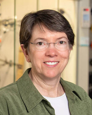
<figcaption>Holly Goodson</figcaption>
</figure>
<p><a href="https://chemistry.nd.edu/faculty/holly-goodson/">Holly Goodson</a>, Professor of Chemistry &amp; Biochemistry, has been selected as a Fellow of The American Society for Cell Biology (ASCB).</p>
<p>She joins 18 other distinguished scientists from across the globe in the 2023 cohort of fellows. Her formal recognition will take place in Boston later this year at Cell Bio 2023, the joint meeting of the ASCB and the European Molecular Biology Organization (EMBO).</p>
<p>"I'm honored to be included in this notable group of scientists. I have long been inspired by the combined breadth and depth of both scientific questions and perspectives exhibited through the ASCB,” she said.</p>
<p>Goodson’s research centers on biological self-organization, focusing on the microtubule cytoskeleton. The cytoskeleton is the structure that helps cells maintain their shape and internal organization, while also providing mechanical support, and is composed of multiple types of filamentous proteins, the largest of which are microtubules.</p>
<p>Microtubules organize the cell’s cytoplasm, among other tasks. Goodson uses a multifaceted approach to determine how the microtubules assemble, what governs their turnover, and how they interact with other parts of the cell. One of the key areas of focus in her lab is the development, alongside collaborators in applied mathematics, of agent-based computational models. These models have allowed Goodson to make connections that experiments alone cannot show because of the number of microtubules.</p>
<p>Beyond research, Goodson serves as science director of the <a href="https://glynnhonors.nd.edu/">Glynn Family Honors Program</a>, and as chair of the ASCB public policy committee. She also broadly serves the cell biology community through educational initiatives, such as co-founding and leading the <a href="https://ibms.nd.edu/">Integrated Biomedical Sciences</a> Ph.D. program at Notre Dame, co-directing an NIH funded post-baccalaureate program (<a href="https://graduateschool.nd.edu/degree-programs/nd-prep-prep---grad-school-prep/">ND-PREP</a>) to prepare researchers from under-represented groups for biomedical Ph.D. programs.</p>
<p class="attribution">Originally published by <span class="rel-author">Rebecca Hicks</span> at <span class="rel-source"><a href="https://chemistry.nd.edu/news/holly-goodson-elected-as-fellow-of-the-american-society-for-cell-biology/">chemistry.nd.edu</a></span> on <span class="rel-pubdate">August 23, 2023</span>.</p>]]>
    </content>
    <link rel="enclosure" type="image/jpeg" href="https://qbio.nd.edu/assets/582658/goodsonnews.jpg" title="Portrait of Holly Goodson"/>
    <author>
      <name>Rebecca Hicks</name>
    </author>
  </entry>
</feed>